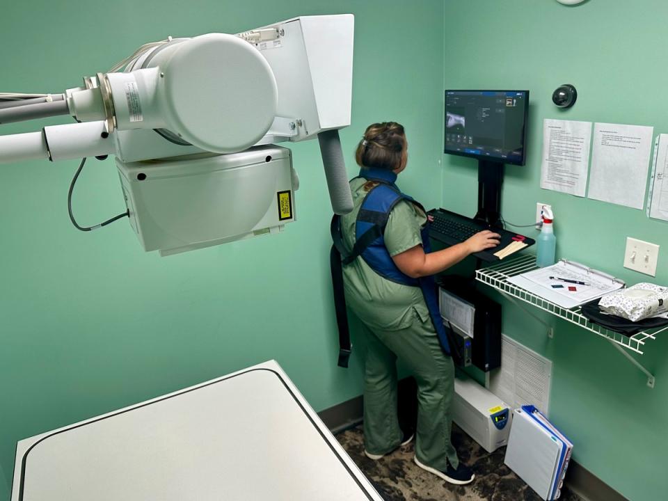
In veterinary medicine, X-rays, also known as radiographs, are a highly useful diagnostic tool we offer at Pine Hollow Veterinary Services. As our patients cannot tell us what they feel or where it hurts, we can take a deeper look at your pet’s organs with X-rays. We are able to diagnose a multitude of problems for our patients including:
- heart disease
- lung disease such as pneumonia
- bowel obstructions
- foreign objects in the digestive tract
- bone fractures, dislocations, or signs of cancer
- arthritis
- the sizes of certain organs, including the heart, liver, spleen, kidneys, urinary bladder, and GI tract
X-rays are like a picture of the inside of your pet. X-rays are painless and noninvasive, meaning that an incision does not have to be made to produce the image. X-rays do not alter the disease process or cause discomfort to your dog or cat. If your pet is in pain or uncomfortable, it may be necessary to use sedation in order to get a clear image.
How does an X-ray work?
An X-ray works by passing electrical energy through the animal, which is absorbed by the tissues. Denser tissues, like bones, absorb more energy and appear whiter on the screen, while less dense tissues, like the liver and spleen, appear gray. Air appears black because it has no density and absorbs no energy. X-rays are produced using the same processes as in human medicine, but the equipment is sized for use in animals.
