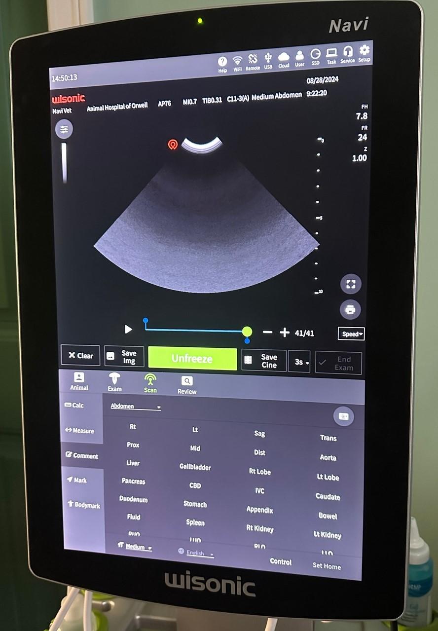
Ultrasound is another non-invasive diagnostic tool available to our patients at Pine Hollow Veterinary Services. Ultrasound is a real-time two-dimensional visualization of the organ systems within the abdomen. It is more sensitive than radiographs allowing visualization of the internal structures of the organs.
During an ultrasound examination of the abdomen, we are looking for:
- architectural or anatomical changes to the organs
- evidence of masses or tumors
- enlarged lymph nodes
- the presence of fluid or gas within the abdominal cavity
- evidence of obstruction of tubular structures such as the common bile duct or ureters
- stones within the urinary bladder or other parts of the renal system
- signs of changes within the organ systems that may be related to the development of disease processes.
How does ultrasound equipment work?
Ultrasound equipment uses sound waves to look at the size and motion of all abdominal organs. Our trained staff will place a probe on your pet’s body and a narrow beam of high-frequency sound waves is directed into the area of interest. The sound waves may be transmitted through, reflected, or absorbed by the tissues they encounter. Images of body structures based on the pattern of echoes reflected from the tissues and organs being imaged are then viewed on a monitor by our staff. Furthermore, an echocardiogram is an ultrasound of the heart to check for wall thickenings and for abnormal valve movements.
Ultrasound imaging can be performed quickly, using minimal restraint or sedation.
To improve the image quality, the area of your pet’s body being scanned is usually shaved, and then ultrasound gel is applied. If we feel that your pet would benefit from having an ultrasound we will perform this procedure right at our clinic during your pet’s examination time. If your pet requires sedation for ultrasound we will accommodate your pet’s needs for the diagnostic procedure.